
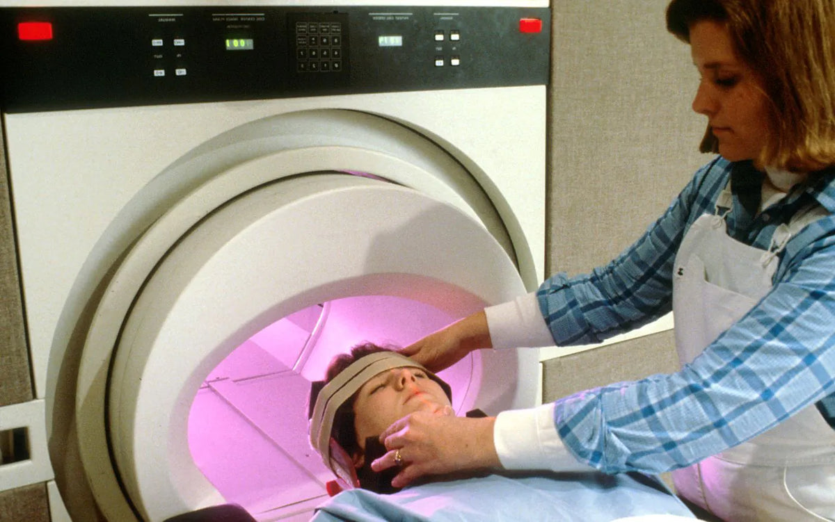
Your brain is burning sugar right now, in the moment you read this sentence. In five healthy men that burning was caught live: glucose with a heavier carbon runs through their veins, and a powerful magnetic scanner follows its signal straight through the skull.
In the recording, one peak splits into two. The small split is the proof: carbon atoms from the infused glucose passed through one exact reaction in the living brain and entered its energy chain.
To leave that trace, the scientists used glucose in which the usual carbon-12 had been replaced with carbon-13. The two isotopes take part in the same chemistry, but the nucleus of carbon-13 has one neutron more and responds to a strong magnetic field in a measurable way. The label [U-13C] says one more important thing: all six carbon atoms in the glucose molecule are labeled. Chemistry can move them around and split them apart, but their magnetic signature stays recognizable.
Five healthy adult men received this glucose through a peripheral venous line. Within the two-hour protocol, 8 grams went in slowly first, then the infusion continued at 8 grams per hour. In three of them it ran outside the magnet and the spectra were recorded after it ended; in two the infusion continued during the scan itself. By the end of the two hours, roughly half the glucose in the plasma carried carbon-13. The 7-tesla system sent radiofrequency pulses and picked up the response of the labeled nuclei from the occipital region of the head as a row of peaks. The position of each depends on the molecule and on the exact place the carbon takes up in it.
One of those peaks belongs to the fifth carbon atom in glutamate. Its splitting shows that a second labeled carbon stands next to it. It is this pair that lets the researchers read the earlier step: the glucose became pyruvate, and part of the pyruvate passed through the pyruvate dehydrogenase complex and entered the tricarboxylic acid cycle as labeled acetyl-coenzyme A. What has to happen between the six heavy carbons in the vein and the two neighboring heavy carbons in glutamate?
The double peak that gives the reaction away
Magnetic resonance spectroscopy tells carbon nuclei apart by their surroundings. Carbon in bicarbonate gives a signal at one place in the spectrum; carbon in a particular position of glutamate gives a signal at another. When two neighboring atoms are both carbon-13, their magnetic states influence each other. The signal from one no longer appears as a single peak, but as a pair. Spectroscopists call that pair a doublet. In the experiment, the doublet D45 links carbon number 5 of glutamate with a labeled carbon number 4 next to it.
A small amount of carbon-13 is present naturally, with no infusion at all. So every measurement comes with a control spectrum recorded without infusion: in the paper it is drawn in blue, and the measurement after the labeled glucose is in red. At the same settings the control gives no recognizable natural signals at the places of bicarbonate and of the fifth carbon of glutamate. After the labeled glucose the red peaks appear exactly there. The splitting adds more than the presence of heavy carbon on its own. One peak would show one labeled nucleus; the doublet shows two neighboring nuclei that stayed side by side through the earlier chemical handovers.
The path to that pair starts before the cycle. Six-carbon glucose splits into two three-carbon pyruvate molecules. Then one carbon leaves each pyruvate as carbon dioxide, and the remaining two-carbon fragment is handed to coenzyme A. The resulting acetyl-coenzyme A joins a four-carbon molecule at the start of the tricarboxylic acid cycle. After a few rearrangements alpha-ketoglutarate forms, and on taking up an amino group it becomes glutamate. So the two neighboring carbons that entered as an acetyl group appear side by side in positions 4 and 5 of glutamate.
The spectrum also holds a high signal from labeled bicarbonate at around 160.8 parts per million. The authors use it as a general trace of the cycle at work, because the carbon dioxide released in the oxidation of glucose quickly equilibrates with bicarbonate. The signal is found in all five volunteers, and in the measurements after the infusion it stays stable for at least an hour. The doublet gives the narrower answer. Its two peaks show that the labeled carbon reached the cycle by the route through pyruvate dehydrogenase.
In 2024 another team measured the composition and reconstructed the architecture of the human pyruvate dehydrogenase complex. A large protein core organizes many copies of three catalytic enzymes and mobile carriers between them.
The machine at the entrance to energy
The core of the complex is built from 60 protein units. Forty-eight carry catalytic sites and bind the enzyme that takes up pyruvate. The remaining twelve anchor the enzyme that collects the electrons at the end of the reaction. When every place on the core is taken, up to 48 copies of the first enzyme and 12 pairs of the last can arrange themselves around it. Together with the helper molecules, the fully loaded assembly reaches about 12 million daltons.
These numbers come from several kinds of measurement. The team separated and weighed the protein parts with mass spectrometry, compared the amounts of the two kinds of unit in gels, added the two outer enzymes until their places were full, and looked at the cores with electron microscopy. The ratio of 48 to 12 holds whether the researchers measure mass, binding, enzyme activity or an electron microscope image.
The reaction has several steps in sequence, and the intermediates have to get from one active site to the next. The large core holds many copies of the enzymes together, and out of its catalytic units run long flexible stretches ending in lipoyl domains. At the end of each one sits lipoamide: a small molecule with a ring holding two sulfur atoms. The flexible link lets this end enter each of the three enzymes in turn. The cryo-electron microscope sees the core far more clearly than these moving parts, because the lipoyl arms keep changing position. That mobility is what lets them carry the intermediates from one active site to the next.
Four vitamins, four work stations
Four B vitamins meet in one and the same machine, but at four different places.
The first meeting is with the enzyme that takes up pyruvate. In its active site sits thiamine diphosphate, a helper molecule made from vitamin B1. When pyruvate binds there, one of its three carbons leaves as carbon dioxide. What is left is a two-carbon fragment with an unstable distribution of electrons. Thiamine diphosphate forms a temporary bond with it and spreads the charge across its own ring. Here the B1 derivative holds the reactive fragment long enough for it to be passed on.
Then the lipoyl arm comes up to the same active site. Its sulfur ring takes the two-carbon fragment from thiamine diphosphate. In the handover the fragment is oxidized to an acetyl group, and the bond between the two sulfur atoms opens and takes up the released electrons. One movement does two jobs: it carries the carbon and it holds the electrons for a while. Lipoamide has its own irreplaceable job among the vitamin derivatives: it carries the two-carbon fragment and the electrons to the next active site.
The loaded arm returns to the catalytic site in the core. Coenzyme A arrives there. Pantothenic acid, vitamin B5, is built into the middle of this long molecule; at one end of it there is a reactive sulfur. It is exactly this end that takes the acetyl group from lipoamide. The new product is acetyl-coenzyme A, which leaves the complex and enters the tricarboxylic acid cycle. The job of B5 is to be part of the molecule that takes the finished two-carbon load and carries it to the next system.
After the handover, lipoamide has to return to its starting state. Its two sulfur atoms now carry the electrons taken up earlier, and they have to form a bond with each other again. This happens in the last enzyme. Fixed inside it is flavin adenine dinucleotide, or FAD, whose reactive part comes from riboflavin, vitamin B2. FAD takes the electrons and so lets the sulfur ring of lipoamide close. Then another helper molecule comes, whose reactive part is built from niacin, vitamin B3. It takes the electrons from FAD and leaves the complex as NADH. FAD stays fixed in the enzyme.
The four vitamins take up four different physical places in one reaction. The B1 derivative holds the carbon fragment. The molecule with B5 takes it and carries it out as acetyl-coenzyme A. The B2 derivative restores the moving lipoyl arm, and the molecule from B3 carries the electrons away as NADH. Acetyl-coenzyme A goes on with the carbon toward the cycle and toward the doublet in glutamate. NADH sets off for the respiratory chain.
How the electrons become usable energy
The carbon trail from the human experiment reaches pyruvate dehydrogenase, the cycle and the products that show up in the spectrum. The road to ATP is taken by the other product of the complex: NADH, which carries the electrons to the respiratory chain. In the inner mitochondrial membrane NADH hands them to the first protein complex of the chain. The electrons pass through further carriers and finally reach oxygen, which together with protons forms water. Along this route the released energy moves protons from the matrix to the space on the other side of the membrane.
The two sides of the membrane now have a different concentration of protons and a different electric charge. The two differences together make an electrochemical gradient, and part of the energy of the electron flow is stored in it. The protons push to come back, but the lipid layer gives them no free passage. Their main controlled route runs through the membrane part of ATP synthase. There the flow drives a ring of c-subunits. Attached to the ring is a long gamma subunit, which turns with it like a shaft.
Above the membrane stands the head of the machine: three alpha and three beta subunits arranged in a still cylinder around the turning shaft. The gamma subunit is asymmetric. As it turns, it presses on the three beta subunits one after another and changes the shape of their active sites. One is open and can release ATP. The second binds ADP and inorganic phosphate more loosely. The third holds them tightly and helps ATP form. With a turn of 120 degrees each beta subunit moves into the next state; in one full revolution all three pass through the whole cycle. The mechanism is called the binding change, because the rotation changes the strength with which each site holds the participants in the reaction.
For a long time this rotation was an inference from the chemical measurements and from the asymmetric structure of the shaft. Then a Japanese team made it visible. The researchers fixed the still head of ATP synthase onto a surface and attached a thin filament of actin to the gamma subunit. Under the microscope the filament began to circle around its axis; with more ATP it turned faster. In this isolated experiment the head breaks ATP down and works in the opposite direction, but the movement shows directly the role of the gamma subunit as a turning shaft. In the whole ATP synthase, the proton flow through the membrane part drives the same mechanics toward synthesis.
This movement drives the synthesis of ATP, a molecule the cell can use straight away. ATP binds to protein pumps, motor proteins and enzymes for synthesis. When its reaction is coupled to their work, it can move ions across a membrane, move cell structures or build a new molecule. NADH brings the electrons, and their movement builds the proton gradient. The gradient turns the shaft, which changes the three active sites one after another.
The small doublet shows that the labeled carbon passed through the first machine and entered the beginning of the cycle. On the blue control spectrum that place stays almost flat. After the infusion of the labeled glucose, two red peaks rise from it.
The same chain runs in your head, with the same four participants: the B1 derivative holds the carbon, the molecule with B5 carries it onward, B2 puts the arm back into service, and B3 carries the electrons away. The experiment only made visible what your brain does without stopping.
Sources
- Sergey Cheshkov et al., Oxidation of [U-13C]glucose in the Human Brain at 7T under Steady State Conditions, Magnetic Resonance in Medicine, 2017.
- Rafal Zdanowicz et al., Stoichiometry and architecture of the human pyruvate dehydrogenase complex, Science Advances, 2024.
- Mulchand S. Patel et al., The pyruvate dehydrogenase complexes: structure-based function and regulation, Journal of Biological Chemistry, 2014.
- Hiroyuki Noji et al., Direct observation of the rotation of F1-ATPase, Nature, 1997.
- Nobel Prize Outreach, The Nobel Prize in Chemistry 1997 — press release.
- Nobel Prize Outreach, The Binding Change Mechanism.


